Root Fracture Treatment in Gurugram
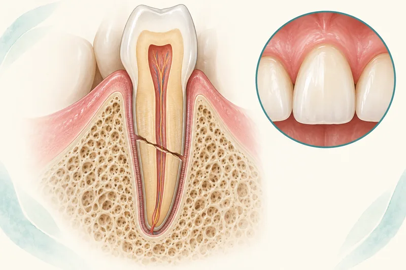
A tooth-root fracture may lie near the gum, in the middle of the root or near its tip. The position and movement of the crown-bearing fragment (the fragment carrying the normally visible tooth part) determine care; the permanent-tooth and baby-tooth pathways are different.
Sources (3)
- Recommended Guidelines for the Treatment of Traumatic Dental Injuries
- International Association of Dental Traumatology guidelines for the management of traumatic dental injuries: 1. Fractures and luxations
- International Association of Dental Traumatology guidelines for traumatic dental injuries: 3. Injuries in the primary dentition
Serious face or jaw injury, uncontrolled heavy mouth bleeding, breathing difficulty from severe swelling, or head-injury signs such as loss of consciousness, vomiting or double vision need emergency medical assessment. Medical care takes priority over preserving the tooth.
Dr. Supreet assesses root-fractured teeth in Gurugram, including tooth type, root development, displacement and pulp status. The pulp is the living tissue inside the tooth; a fracture does not automatically mean that root canal treatment (treatment inside the tooth) or extraction is needed immediately.
Sources (3)
Locating the fracture
The dentist uses the injury history, examination and appropriate pulp tests and dental images. A permanent root fracture may need images from different angles. Cone-beam CT, a three-dimensional dental scan, can be considered if ordinary images leave important treatment-planning questions unanswered; it is selective rather than routine.
Repositioning or monitoring a permanent tooth
A displaced crown-bearing segment in a permanent tooth should be repositioned promptly by the dentist, checked with dental imaging and stabilized with a passive flexible splint (temporary tooth support). The guideline usually describes about four weeks of stabilization; a fracture near the gum, called a cervical fracture, may need up to four months. The injury and movement determine the plan.
A stable crown-bearing fragment should not be removed merely because the fracture is cervical. Cervical fractures can heal. Root canal treatment is not started routinely at the emergency visit: the tooth and pulp are monitored, and an early negative response to pulp testing alone does not establish pulp death.
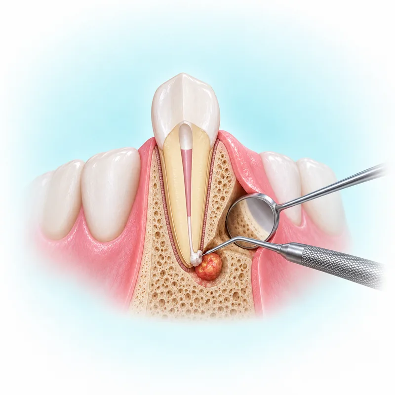
If pulp death and infection later develop in the crown-bearing segment, treatment may be limited to that part rather than the root segment near the tip. This follows established findings during review, not a routine immediate root-canal rule.
Later treatment for selected fractures near the gum line
For a fully developed permanent tooth with a fracture above the bone crest (the upper edge of supporting bone) and a very mobile crown-bearing fragment, later reconstruction may involve removing that loose segment, root canal treatment and a post-retained crown (a protective cap held by a support inside the tooth). Exposing a restorable root by orthodontic or surgical means, or extraction, may be discussed. This narrow later exception does not justify routine removal of a stable cervical fragment at the emergency visit; feasibility and visits require individual assessment.
For children or teenagers with a permanent root-fractured tooth needing stabilization, one hospital patient pathway describes a thin flexible wire and cement bonded to the injured and neighbouring teeth, with visits to check and remove the splint. It may remain for several weeks and, for some root fractures, up to about four months. That appearance and care description does not establish an adult or baby-tooth procedure or one duration for everyone.
Baby-tooth root fractures
These baby-tooth examples come from guidance for otherwise medically healthy children with previously sound, decay-free baby teeth. Medical problems, tooth decay or other injuries need their own assessment.
A baby-tooth root fracture may be observed when the crown-bearing fragment is not displaced or excessively mobile. If it is displaced, very mobile and interfering with the bite, a child-oriented team may consider fragment removal or repositioning with selective flexible splinting. Displacement, mobility, bite and cooperation determine the choice; the permanent default repositioning rule cannot simply be copied.
Baby-tooth follow-up differs after observation, repositioning and splinting, or fragment removal. The developing permanent tooth also needs consideration. Parents should return for gum swelling, a draining gum lesion, increasing mobility or other concerns; color change alone is not always infection.
Care and review after treatment
Follow the treating team's gentle-brushing, soft-food and injured-tooth biting instructions after trauma treatment, with contact-sport restrictions as advised. If a splint was placed and becomes loose, arrange dental review. A new gum lump or facial swelling needs reassessment; uncontrolled bleeding or severe swelling needs urgent assessment. These signs do not establish the complication by themselves.
Clinical and dental-image checks monitor healing and pulp status after repositioning or splinting, because complications can develop later. Relevant sports mouthguards reduce future trauma risk but cannot eliminate it.
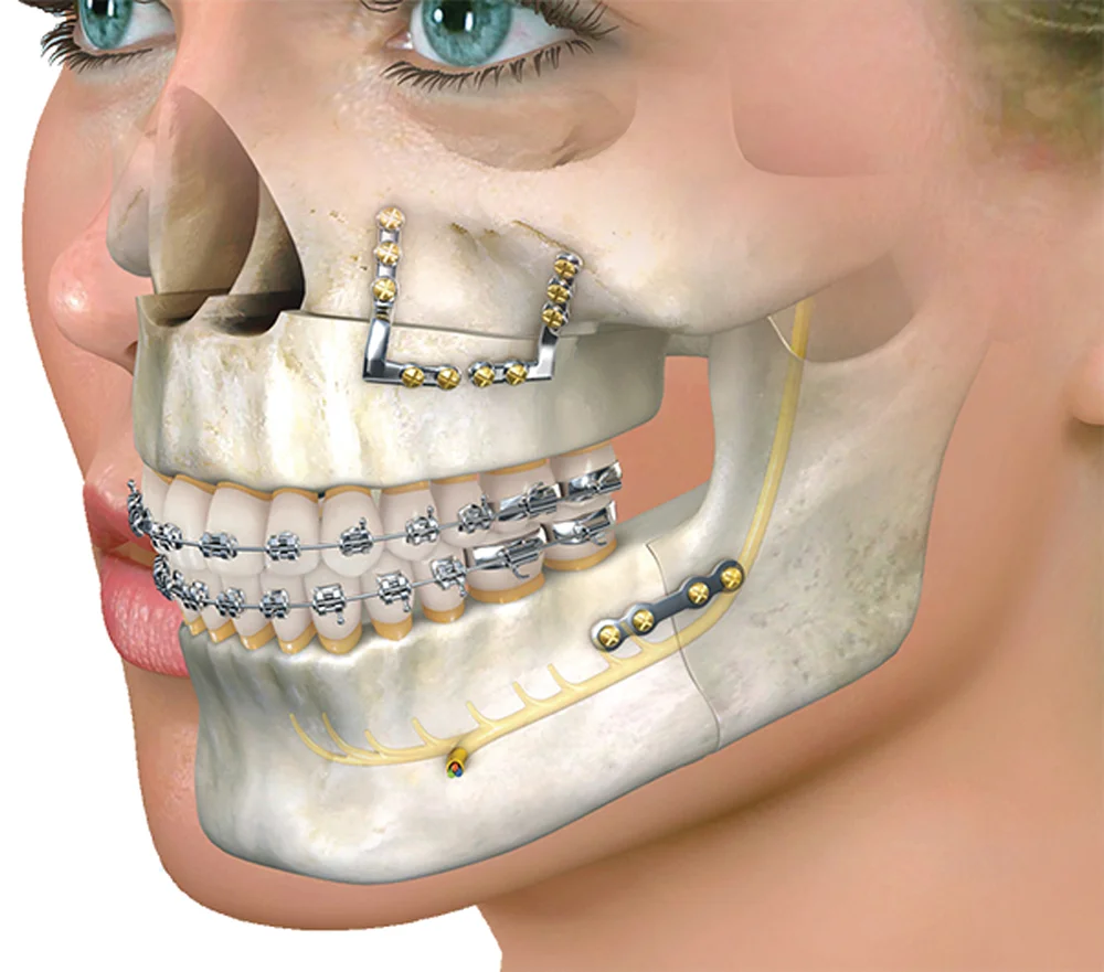
For the wider specialist assessment, see oral and maxillofacial surgery.
Arrange assessment in Gurgaon
Dr. Supreet Kaur Sawhney, MDS, Senior Consultant – Oral & Maxillofacial Surgeon and Senior Implantologist, brings 20+ years of experience to assessment and individual treatment planning. Consult at Dr. Supreet's Dental & Implant Centre, 395 P, Sector 39, near Medanta Hospital, Gurugram (Gurgaon), Haryana 122001. Contact the clinic, call +91 9650298009, or view the Sector 39 clinic. Medical danger signs need emergency assessment; do not wait for a routine appointment.