Displaced Permanent Tooth Treatment in Gurugram
A permanent tooth displaced by injury may move partly out of its socket, sideways, or deeper into the socket. These are different injuries: extrusion, lateral luxation and intrusion. The dentist must identify the direction, root development and any associated bone damage before selecting treatment.
Sources (3)
Serious face or jaw injury, uncontrolled heavy mouth bleeding, breathing difficulty from severe swelling, or head-injury signs such as loss of consciousness, vomiting or double vision need emergency medical assessment. Medical care takes priority over tooth preservation.
Dr. Supreet assesses and treats displaced permanent teeth in Gurugram. Seek prompt dental assessment after displacement rather than selecting a repositioning method yourself. This guide concerns permanent teeth; baby-tooth protocols are different and must not be inferred from these pathways.
Sources (2)
Assessment and pulp tests
The dentist may use injury history, examination, mobility, tapping, pulp tests and dental images according to the injury. The pulp is the living tissue inside the tooth. An absent response to an early pulp test soon after injury does not prove pulp death; serial clinical and dental-image findings are important.
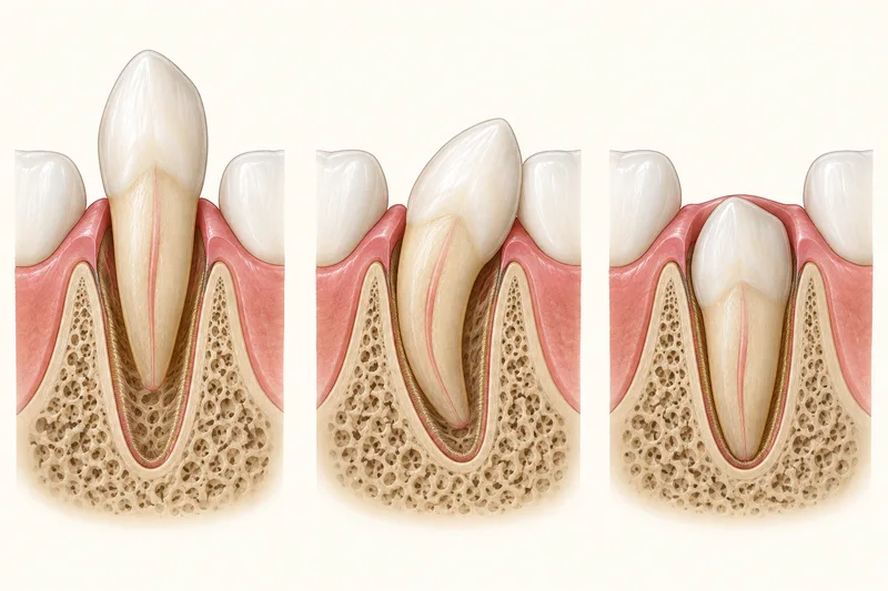
Extrusion: partly out of the socket
An extruded permanent tooth may look longer because it has moved partly out. The IADT pathway describes gentle clinician repositioning under local anaesthesia (numbing the treatment area) and a passive flexible splint for two weeks. A fracture or breakdown of the marginal bone (bone around the socket edge) can require four additional weeks. A splint is temporary support, and associated injury determines the actual plan.
Sources (2)
The pulp needs monitoring after extrusion. If pulp death and infection are established, treatment is chosen for the root's development stage; one early negative pulp test alone is not an indication for root canal treatment (treatment inside the tooth). Clinical and dental-image checks begin in the early weeks and may continue with yearly reviews for at least five years, adapted to findings and associated injury.
Lateral luxation: sideways or angled displacement
A laterally displaced permanent tooth can feel firm because it may be locked in a damaged socket; not every displaced tooth is loose. A dentist can disengage and reposition it under local anaesthesia (numbing the treatment area), then use a passive flexible splint for about four weeks. Associated socket or marginal-bone fracture (around the socket edge) can change stabilization.
Sources (2)
Pulp planning after lateral luxation differs with root maturity. An incompletely developed permanent tooth may regain its pulp blood supply; pulp death is more likely with a fully developed root and may require treatment inside the tooth. Clinical and dental-image assessment guide timing; an absent response to an early pulp test alone does not diagnose pulp death. Repeated checks continue through at least five years because late infection, root breakdown or fusion to surrounding bone can occur.
Intrusion: driven into the socket
An intruded permanent tooth has been driven into its socket and bone. The dentist assesses measured displacement, root maturity, pulp status and associated injury with examination and appropriate dental images. This is not a home measurement or a reason to wait for initial care.
Sources (2)
For an intruded permanent tooth with an incompletely formed root, the IADT pathway initially allows spontaneous re-eruption, movement back toward its normal position. If it has not re-erupted within four weeks, the dentist starts orthodontic repositioning (clinician-directed tooth movement). Pulp monitoring continues; infected pulp death or inflammatory root breakdown may later require treatment suited to the developing root. These are review decisions after professional assessment.
Sources (2)
For a fully formed permanent root, the measured intrusion depth changes the pathway: under 3 mm may initially be observed for re-eruption, with clinician repositioning if it has not returned within eight weeks; 3–7 mm is generally repositioned surgically or orthodontically; over 7 mm is surgically repositioned. The clinician selects the method, stabilization and associated-injury care. These thresholds do not authorize delaying the first assessment.
After intrusion of a fully developed permanent tooth, pulp death is very likely. The IADT pathway calls for root canal treatment (treatment inside the tooth) around two weeks or once position permits access, to reduce infection-related root breakdown. This mature-root pathway does not apply automatically to an immature permanent root or a baby tooth.
The intrusion guideline describes clinical and dental-image review around two, four, eight and twelve weeks, six months, one year and then yearly for at least five years. Re-eruption, pulp infection, root breakdown and fusion to bone are monitored, with care adjusted when adverse findings develop.
Protect healing and report changes
Follow the treating team's injury-specific cleaning, diet, biting and sports instructions after treatment. A placed splint that becomes loose needs dental review. New gum lumps or facial swelling need reassessment, and uncontrolled bleeding or severe swelling needs urgent assessment. After luxation care, report new or worsening discoloration, continuing pain, bleeding or swelling; these signs do not establish a particular complication by themselves.
Sources (3)
Keep the reviews even when the tooth feels comfortable, because later pulp or root complications may occur. Relevant sports mouthguards can reduce future trauma risk but cannot eliminate it. None of these pathways promises survival or the same treatment for every injury.
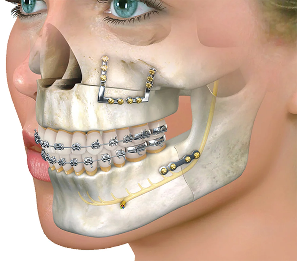
For the wider specialist assessment, see oral and maxillofacial surgery.
Arrange assessment in Gurgaon
Dr. Supreet Kaur Sawhney, MDS, Senior Consultant – Oral & Maxillofacial Surgeon and Senior Implantologist, brings 20+ years of experience to assessment and individual treatment planning. Consult at Dr. Supreet's Dental & Implant Centre, 395 P, Sector 39, near Medanta Hospital, Gurugram (Gurgaon), Haryana 122001. Contact the clinic, call +91 9650298009, or view the Sector 39 clinic. Medical danger signs need emergency assessment; do not wait for a routine appointment.