Laser Gum Depigmentation in Gurugram
Diagnose the pigment before cosmetic treatment
Dr. Supreet Kaur Sawhney in Gurugram assesses the cause of gum pigmentation before elective colour treatment. Melanin is natural pigment, and stable physiologic pigmentation is not itself a disease or automatically a reason for biopsy (taking a tissue sample). A new or enlarging pigmented area needs assessment and possible biopsy; a clinically suspicious adult lesion needs prompt biopsy or specialist referral rather than cosmetic removal without diagnosis.
Sources (3)
The gum surgery and grafting guide explains the broader gum-health consultation before an appearance treatment is planned.
Ablative and nonablative methods differ
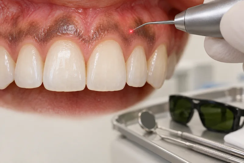
Ablative laser treatment removes the surface gum lining that contains pigment. Nonablative methods target pigment without removing that lining. Devices and mechanisms differ; the same procedure, care or result should not be assumed for every method.
A nonlaser option and limited comparisons
A small study used a blade and a diode laser on different sides of 20 selected patients after initial periodontal care. Pain and early pigment-return differences were not statistically clear during its short three-month follow-up. That does not prove the methods equivalent, a permanent result, or suitability for other pigment causes or devices.
One selected healthy-adult ablation example
In a study with 24 selected healthy adults, eight participants received 980-nm diode laser ablation of the front gum surface after local numbing and wavelength-matched protective eyewear. Smokers, pregnancy or lactation, and systemic or syndromic pigmentation were excluded. These are study details, not a description of clinic equipment, settings or a standard protocol.
In that study, participants avoided hot and spicy food for the first postoperative week and returned for pain and wound-healing checks. Discomfort and healing varied; the small diode group did not establish universal faster healing or pain-free treatment. The food restriction and reviews belong to this selected healthy-adult 980-nm ablation protocol. Follow your surgeon’s actual instructions; these directions do not transfer to nonablative methods.
Pigment may return
A separate trial followed 60 people with physiologic pigment on the upper cheek-side gums for two years after two specified laser treatments. Pigment reappeared at later follow-up. This does not predict recurrence for every person, site or technique, or promise a permanent colour change.
Bleeding after an ablative wound
After an actual ablative gum-depigmentation wound, bleeding that continues despite firm pressure needs contact with the treating dental or surgical team or urgent care. This is general mouth-surgery wound advice used for persistent bleeding, not a laser-specific complication rate or a fixed pressure duration. Your treating team’s instructions take priority; it is not a shared nonablative aftercare plan.
Arrange an assessment
Dr. Supreet Kaur Sawhney, MDS, is a Senior Consultant Oral & Maxillofacial Surgeon and Senior Implantologist with more than 20 years of experience. Dr. Supreet’s Dental & Implant Centre is at 395 P, Sector 39, near Medanta Hospital, Gurugram (Gurgaon), Haryana 122001. For an assessment, contact the clinic or call +91 9650298009.