Impacted Tooth Imaging in Gurugram
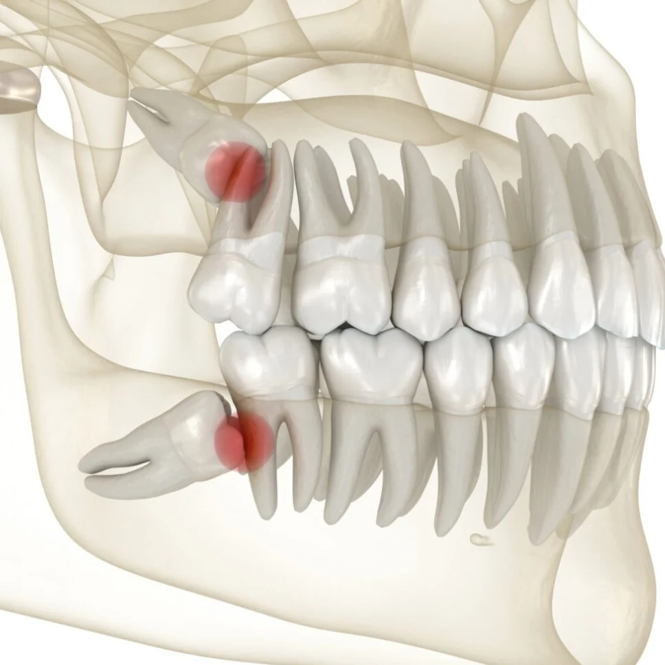
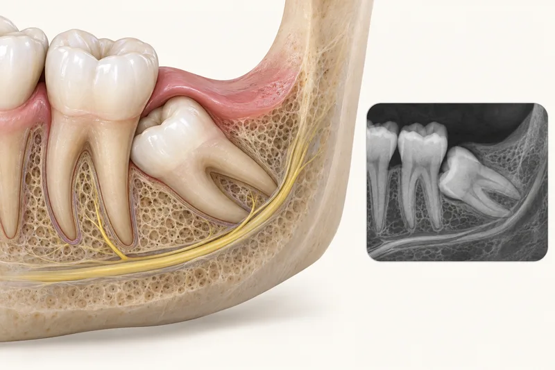
Imaging can help locate an impacted tooth and assess nearby structures. A panoramic dental X-ray provides an overview; selected three-dimensional imaging may be considered when examination and suitable images using less radiation cannot answer a specific planning question. Dr. Supreet Kaur Sawhney, MDS in Oral & Maxillofacial Surgery, is a Senior Consultant and Senior Implantologist with 20+ years of experience.
Sources (3)
Why imaging is selected
A panoramic dental X-ray, taken with equipment outside the mouth, gives an overview of the teeth and jaws in one two-dimensional image and can help assess an impacted tooth. Dental X-rays are selected using the clinical findings, history and disease risk; existing suitable images should be considered before ordering more.
Sources (3)
A panoramic image does not show precise detail for each tooth or soft tissues, such as gums, and is not a three-dimensional scan. When it cannot answer a specific question, targeted or other imaging may be needed.
Sources
Dental X-rays involve radiation. The expected diagnostic benefit should outweigh the exposure risk, and the radiation dose should be kept appropriate for the question being answered.
When further imaging may be considered
For selected complex impacted-tooth surgical planning, dental cone-beam computed tomography (CBCT) may show the tooth and nearby jaw anatomy in three dimensions—but only when examination and images using less radiation do not answer the planning question. It is not routine for every impaction and does not itself prove that surgery is needed.
Sources (2)
For an unerupted ectopic upper canine (an upper pointed tooth that has not come through normally and is in an unusual position), conventional X-rays taken from different angles (parallax views) can first help locate the tooth and assess possible damage to nearby front-tooth roots. If those images answer the planning question, no further image is needed; CBCT of a small selected area may be considered when findings remain uncertain or complicated. This is a selected canine pathway, not a universal protocol for wisdom teeth or adults.
Experience and next steps
For an image taken with equipment outside the mouth, such as a panoramic dental X-ray, the machine rotates around the head and the patient follows instructions to remain still. Setup differs by equipment. For CBCT, the patient sits or stands while the scanner moves around the head; the scan is painless, but the team should know about possible pregnancy or difficulty remaining still.
Sources (2)
The clinician interprets images with the examination and explains whether more imaging or treatment is needed. A scan does not decide by itself between monitoring, orthodontic care or surgery. Seek prompt dental assessment for severe or persistent dental pain or growing mouth swelling. Uncontrolled mouth bleeding or swelling that makes breathing difficult needs emergency medical assessment. These actions concern the underlying dental symptoms; they are not scan effects or instructions to repeat imaging.
Sources (3)
Once the imaging question is answered, compare the options in impacted-tooth assessment and surgery and the wider oral and maxillofacial assessment guide.
Discuss Impacted Tooth Imaging in Gurugram
Dr. Supreet’s consultation can help review findings, alternatives and next steps. Dr. Supreet’s Dental & Implant Centre is at 395 P, Sector 39, near Medanta Hospital, Gurugram (Gurgaon), Haryana 122001. Bring prior records and a current medicine list. View the Sector 39 clinic, contact the clinic or call +91 9650298009 to ask about an appointment.