Chipped and Broken Tooth Treatment in Gurugram
A visible tooth chip may involve only enamel, the hard outer layer, or hide a deeper injury. A crown fracture affects the normally visible part of the natural tooth; the dentist checks whether the layer beneath enamel, called dentin, or the living pulp tissue inside is involved.
After an injury, serious face or jaw damage, uncontrolled heavy mouth bleeding, breathing difficulty from severe swelling, or head-injury signs such as loss of consciousness, vomiting or double vision need emergency medical assessment. Medical care takes priority over repairing a tooth.
Dr. Supreet assesses chipped and fractured teeth in Gurugram, including whether a tooth is baby or permanent, its root development and any displacement or root fracture. A small-looking chip should be checked; symptoms alone do not establish how deep it is.
Sources (2)
Protect the tooth while reaching care
Avoid hard chewing on the injured tooth before assessment. If a fragment has broken off, take it in milk or saliva in a container; this is fragment storage, not advice to replant a baby tooth.
For a crown fracture painful with air or cold drinks, gently biting on clean, moist gauze or cloth may provide temporary relief while reaching the dentist. It is not a repair or proof of pulp exposure. Do not place aspirin or topical oral pain ointment on the area; prompt professional examination is still needed.
Sources
The dentist examines the tooth and may use dental images according to the injury. Tooth mobility, tenderness and pulp testing can suggest an associated displacement or root injury; visible chip size alone cannot exclude it.
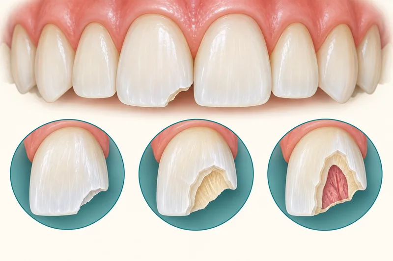
An enamel-only fracture of a permanent tooth
In this branch, enamel is missing but dentin is not exposed. According to the extent and location, a dentist may smooth the edge, rebond a usable fragment or place a composite resin restoration (tooth-coloured repair resin). A deeper fracture or associated displacement or root fracture needs a different plan; these are clinician procedures, not home repairs.
Sources (3)
For an isolated permanent enamel-only fracture, the IADT guideline describes clinical and dental-image review at about six to eight weeks and one year, adjusted to findings. Suspected or confirmed displacement or root fracture requires its longer injury-specific review plan. Pain or temperature sensitivity needs assessment because the injury may extend deeper; those symptoms do not diagnose the depth by themselves.
Sources (2)
A permanent crown fracture involving dentin
If dentin is exposed but the pulp is not, the clinician protects the exposed layer with a suitable restoration. An intact, usable fragment may be reattached; a filling or crown (a protective cap) may be discussed according to the remaining tooth. Associated displacement or root fracture changes the assessment and review plan.
Sources (3)
For selected chips suitable for direct composite bonding, tooth-coloured resin is conditioned, shaped, hardened and polished on the tooth. Bonding is not treatment for every deeper fracture or painful tooth. The material can stain or chip; persistent roughness or an abnormal bite after bonding needs reassessment. Daily cleaning and avoiding hard-object biting help maintain it, alongside dental reviews.
Sources
When a permanent crown fracture exposes pulp
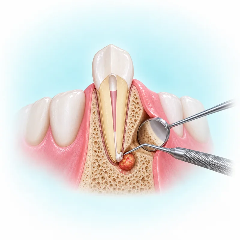
Pulp capping or partial pulpotomy, protecting the pulp or removing part of the injured pulp tissue, may preserve viable tissue. This matters especially when a permanent root is still developing, but conservative pulp care can also suit many fully developed teeth. A tooth with a fully developed root that needs a post (a support to retain a protective crown cap) may instead need root canal treatment (treatment inside the tooth). Pulp condition, remaining tooth, root maturity and associated injury determine the choice; pulp exposure does not make root canal treatment automatic.
Sources (3)
After appropriate pulp care, a usable fragment may be moistened and bonded back, or the tooth restored with another suitable material. The sequence depends on the injury and remaining tooth; a one-visit repair is not promised. Later problems can include discoloration, pulp death or infection, pain around the root, or restoration loss or breakdown. These are outcomes to monitor, not a prediction that they will occur.
The permanent enamel-dentin fracture guideline describes review around six to eight weeks and one year without pulp exposure, with additional three- and six-month reviews for pulp-exposing fractures. If displacement or root fracture is also present, its schedule takes precedence and may continue longer. The dentist arranges the actual clinical and dental-image follow-up.
Baby-tooth fractures need their own choices
These baby-tooth examples come from guidance for otherwise medically healthy children with previously sound, decay-free baby teeth. Medical problems, tooth decay or other injuries need their own assessment.
For an isolated enamel-only chip in a baby tooth, a clinician may smooth a sharp edge; the primary-tooth guideline does not recommend routine clinical or dental-image follow-up for that isolated injury. Dentin, pulp, displacement, soft-tissue or root injury changes this advice. Do not apply the permanent enamel-fracture schedule automatically to a baby tooth.
For a baby crown fracture exposing dentin without pulp exposure, the dentist covers the layer with a suitable material and the guideline advises a clinical check around six to eight weeks. With exposed pulp, a child-oriented team may consider partial pulpotomy under local anaesthesia (numbing the treatment area), discussing the child's maturity, ability to tolerate care and the exposure with parents. Treatment or rapid referral follows the individual assessment.
For primary pulp-exposing crown fractures, the guideline describes clinical reviews around one week, six to eight weeks and one year, with imaging and further treatment depending on findings and pulp care. Parents should return earlier for gum swelling, a draining gum lesion, increasing mobility or other concerns. A color change alone is not always infection.
Care after treatment
Follow the team's gentle-brushing, soft-food and injured-tooth biting advice after trauma treatment. Avoid contact sports while healing as instructed. If a splint (temporary tooth support) was placed and becomes loose, contact the dental team; a new gum lump or facial swelling needs reassessment, while uncontrolled bleeding or severe swelling needs urgent assessment. These signs do not diagnose a complication by themselves. Sports mouthguards may reduce future injury risk without eliminating it.
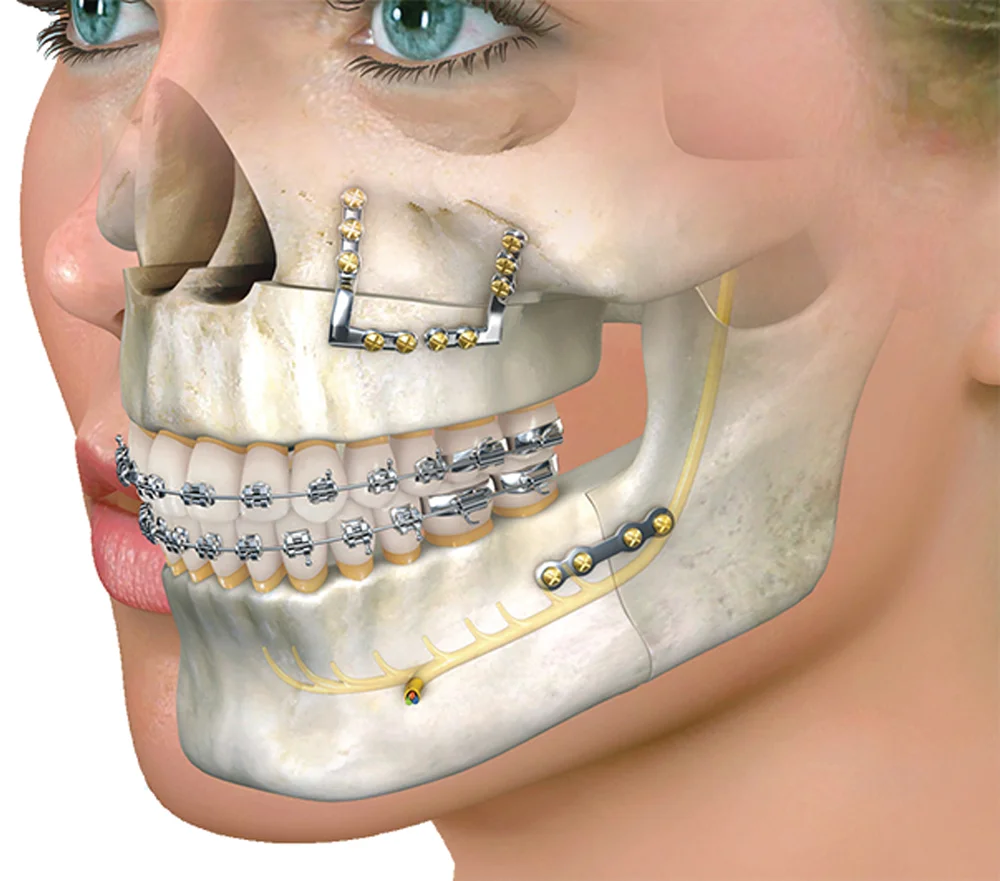
For the wider specialist assessment, see oral and maxillofacial surgery.
Arrange assessment in Gurgaon
Dr. Supreet Kaur Sawhney, MDS, Senior Consultant – Oral & Maxillofacial Surgeon and Senior Implantologist, brings 20+ years of experience to assessment and individual treatment planning. Consult at Dr. Supreet's Dental & Implant Centre, 395 P, Sector 39, near Medanta Hospital, Gurugram (Gurgaon), Haryana 122001. Contact the clinic, call +91 9650298009, or view the Sector 39 clinic. Medical danger signs need emergency assessment; do not wait for a routine appointment.