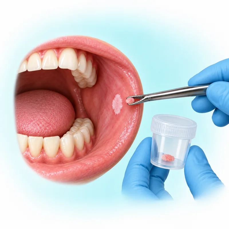
Hard-Tissue (Jawbone) Biopsy in Gurugram
A hard-tissue biopsy takes a sample of jawbone or an abnormal area within it for examination under a microscope, called histopathology. It is a diagnostic procedure: an unusual finding on an X-ray does not by itself settle the diagnosis or mean that a biopsy is always needed. Dr. Supreet Kaur Sawhney provides jawbone-biopsy care in Gurugram, with assessment of the reason for sampling, the appropriate approach and the result pathway.
Sources (2)
When might a jawbone sample be considered?
An unexplained or expanding jawbone lesion, meaning an abnormal area, may need biopsy. So may a periapical abnormality, a change around a tooth’s root tip, that does not resolve. The examination, possible causes and imaging guide the decision; not every jawbone X-ray finding needs the same procedure.
Sources
Dr. Supreet holds an MDS in Oral & Maxillofacial Surgery and has 20+ years of experience. She is a Senior Consultant – Oral & Maxillofacial Surgeon and Senior Implantologist. Bring your referral, previous reports and existing images to Dr. Supreet's Dental & Implant Centre in Sector 39 so the diagnostic question can be discussed.
A jawbone biopsy samples bone or tissue inside it. The oral-biopsy guide discusses sampling the fleshy mouth lining and choosing partial or whole-lesion sampling. Bone sampling for diagnosis has a different purpose from reshaping bone for a denture, treating a tooth’s root end or draining a dental abscess. The clinical findings determine which question needs to be addressed.
Sources
Choosing images to answer the clinical question
X-rays help locate a lesion inside the jawbone and plan where to sample. Images are interpreted alongside the clinical examination; an image alone does not provide every part of a diagnosis or show every nearby structure. The choice depends on the findings, history and individual disease risk, rather than an automatic imaging schedule at every visit.
Sources (2)
Suitable existing images should be considered first. Further images may be needed if the earlier views cannot answer the question. Dental X-rays use ionizing radiation, so the expected diagnostic benefit should outweigh the exposure risk and the imaging should use an appropriate, optimized dose.
Sources
Cone-beam computed tomography (CBCT), a three-dimensional dental X-ray scan, should be selected only when the clinical question cannot be answered adequately with lower-exposure imaging after examination. It is not a routine replacement for two-dimensional X-rays or a necessary scan for every jawbone biopsy.
Sources
How the sampling approach is chosen
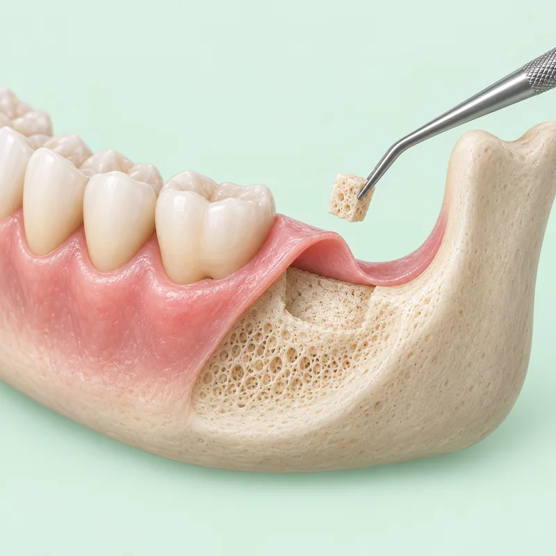
Jawbone sampling may use an open approach or a needle technique. The diagnostic problem and jaw anatomy determine the selection. Discuss what tissue needs to be obtained, how it can be reached and the risks of the proposed approach before deciding on the plan.
Sources
For an open intraoral biopsy, meaning an approach through the mouth, the surgeon makes a small cut in the gum, obtains a bone or other tissue sample from within the jawbone and may use stitches. The approach is planned to avoid nearby important structures and remove only the bone needed to reach the target. That planning reduces unnecessary bone removal; it does not guarantee that nerves or nearby tissues cannot be injured. This describes the open route, rather than every needle biopsy.
If the features suggest that the abnormal area may contain blood or fluid, aspiration—drawing out fluid with a needle—may be needed before tissue sampling. A possible vascular lesion, involving blood vessels, needs particular specialist caution. Aspiration is not an automatic step for every jawbone lesion.
Sources
Risks and care depend on the site and approach
Possible complications include bleeding, infection, nerve injury and harm to nearby structures. The likelihood and consequences vary with the lesion’s location, jaw anatomy and the approach, so the consent discussion should address the proposed procedure. No single risk rate applies to all jawbone biopsies.
Sources
After an open biopsy through a gum incision, avoid biting numb mouth tissue and avoid vigorous rinsing on the procedure day. Follow the treating team’s cleaning and food advice. These oral-wound instructions concern the open approach; care after a needle biopsy follows its individual procedural plan.
If an open gum-incision biopsy wound bleeds, apply firm pressure as instructed and contact the treating team promptly if it does not stop. Persistent or heavy bleeding needs urgent local clinical assessment; do not wait for the biopsy result.
How you will receive your biopsy result
The team should arrange to communicate the microscopic tissue report, either at a review or in writing. A return appointment is not automatically necessary for every patient. Further assessment or treatment depends on the diagnosis; taking a sample does not promise a particular result or complete every treatment. Before leaving, understand how and from whom the result will be communicated.
Discuss jawbone-biopsy care in Gurgaon
Dr. Supreet's Dental & Implant Centre is at 395 P, Sector 39, Near Medanta Hospital, Gurugram, Haryana 122001. A consultation can address the reason for sampling, existing images, the proposed route, site-specific risks and the result plan. The broader oral and maxillofacial surgery guide explains how findings guide treatment or referral. An estimate follows the individual plan.